Prof. Kenro Kusumi (R) with authors of the paper, Jeanne Wilson-Rawls (L) and Elizabeth Hutchins (centre). Photo credit: Joel Robertson.
We, continue our Researcher of the Month initiative, with an interview with Professor Kenro Kusumi, who studies development, regeneration and diseases of the spine in his lab at the School of Life Sciences at the Arizona State University. Prof. Kusumi’s expertise lies in developmental biology, embryology, evolution and genomics and recently published a paper in PLOS ONE on his findings from tail regeneration seen in green anole lizards which will pave way to finding regenerative treatment methods for diseases such as arthritis, scoliosis etc.
CTS: For the benefit of our readers, could you please summarize your recent findings?
KK: In order to examine the genes that are differentially expressed within the regenerating lizard tail, we used RNA-Seq to assess all the genes expressed at 25 days of regeneration. This is a stage that marks maximal growth of the lizard tail, with formation of new tissues towards the base and patterning of those tissues towards the tip. We were able to read all the output of the 23,000 or so lizard genes in five sections along the tail in multiple replicates (n=5). By carrying out bioinformatic analysis and statistical tests, we found that at least 326 genes were turned on in specific spots within the regenerating tail. This gave us the first clues in the lizard as to the genetic ‘recipe’ for regeneration, and where each of the ingredients has to be used.
CTS: How do you envisage that your findings will be applied to treating patients in the future.

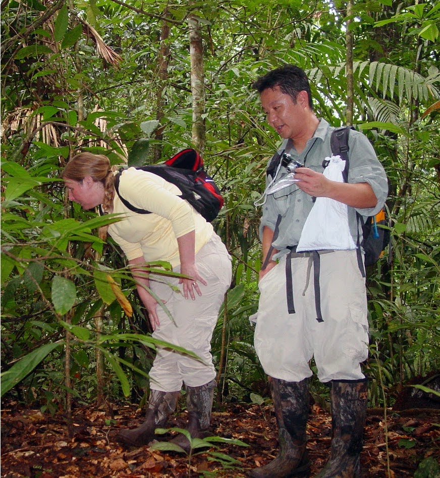
Prof. Kusumi with Prof. Jeanne collecting specimens of anole lizards in Panama (Photo courtesy: Kenro Kusumi)
KK: Regenerating major parts of the human body parts will be a challenge for the future. This research points out some of the avenues towards that goal. As anyone who suffers from arthritis knows, an important part of the limb is the joints, which are cushioned by a specific type of cartilage. Lizards grow lots of this cartilage in their regenerated tails, and we hope that this process can be activated to repair arthritis in humans. Lizards also regrow their spinal cord and their ability to use their tails. Activating nerve regeneration could help people suffering from spinal cord injury and would be necessary to regrow limbs.
Because lizards and people have a relatively recent common ancestor, compared with frogs and fish, we share many more similarities in the genome or the genetic instructions within each cell. Almost all of the 326 genes that we identified in the regeneration are shared between lizards and humans. Over time, we suspect that these genes have changed between lizards and humans as to where and how much they are expressed in the tissue after injury. There are already drugs developed that alter the expression of some of these families of genes, and our hope is that in the future, we can reprogram cells for regenerative therapies.
CTS: Since we have a fair idea of how regeneration works and like you mentioned, even have a few drugs that can alter expression of certain genes, how soon would you reckon, that regenerative medicine will become part of mainstream treatments?
KK:This is a hard question to answer, since clinical trials can be very time-consuming and slow. I would hope that findings within the next 5 years could then find applications in the clinic and be fully tested in the next 20 years.
CTS: What are the greatest hurdles that these treatments need to pass before they become every day techniques.
KK: Testing in mammalian model systems will be important. While genes that regulate regeneration and cell proliferation can be activated, it will be critical to establish the safety of activating genetic pathways in models like to mouse, to avoid any unintended consequences.
CTS: Mice tend be to be preferred choice of models when it comes for experimentation and there is evidence for regeneration in them too. Why have you chosen green anole lizards as your model organism?

Anolis carolinensis (Photo credit: Wikipedia)
KK: While mammals display limited regenerative capacity, such as the digit tips at neonatal stages (Rinkevich et al., 2011), lizards are the most closely related animals to humans that are able to regrow entire body appendages as adults, such as the tail that includes tissues such as skin, skeletal muscle, cartilage, blood vessels, and spinal cord. This capacity for regeneration is also observed in fish (e.g., zebrafish) and amphibians (e.g., axolotl, newt, and frog tadpoles), but not in mammals and birds. The genome sequence of the first lizard, the green anole, was published in 2011 (Alföldi et al., Nature) and molecular analysis of the process in the lizard model became possible. Building on this genome with high coverage annotation (Eckalbar et al., BMC Genomics, 2013), we were able to identify the genes that are differentially expressed along the growing lizard tail.
CTS: Why is it that blastemas do not work in more developed organisms such as mice and humans, when they seem to working well in lesser developed animals such salamanders.

Ms. Hutchins enjoying a lighter moment during her field trip in Panama. (Photo courtesy: Kenro Kusumi)
One of the key findings of this paper is that regeneration of the lizard tail appears to follow a non-blastema, more distributed model. Evidence for this comes for two major sources: distribution of markers for proliferating cells (PCNA and MCM2O and patterns of gene expression of stem/progenitor cells markers within the lizard tail. In the regeneration of the newt, PNCA and MCM2 immunostaining localizes regions of proliferative growth in the blastema. In contrast, in the lizard tail, PCNA and MCM2 immunostaining is observed is regions of tissue all along the regenerating tail, and skeletal muscle and cartilage differentiation occurs along the length of the regenerating tail during outgrowth. It is not limited to the most proximal regions. Furthermore, the distal tip region of the regenerating lizard tail is highly vascular, unlike a blastema, which is avascular. Genes for stem cell and progenitor cells (hematopoietic, musculoskeletal) are highly expressed in purified lizard satellite cells or embryos, but there is no region of elevated expression within the regenerating tail. Together, these data suggest that the blastema model of anamniote limb regeneration does not accurately reflect the regenerative process in tail regeneration of the lizard, an amniote vertebrate. That is relevant for developing human therapies; if a blastema is not a conserved feature of the regenerative process in amniotes, then we would be pursuing the wrong direction in trying to recreate one in mammals.
References:
Eckalbar WL, Hutchins ED, Markov GJ, Allen AN, Corneveaux JJ, Lindblad-Toh K, Di Palma F, Alföldi J, Huentelman MJ, & Kusumi K (2013). Genome reannotation of the lizard Anolis carolinensis based on 14 adult and embryonic deep transcriptomes. BMC genomics, 14 PMID: 23343042
Rinkevich Y, Lindau P, Ueno H, Longaker MT, & Weissman IL (2011). Germ-layer and lineage-restricted stem/progenitors regenerate the mouse digit tip. Nature, 476 (7361), 409-13 PMID: 21866153
Hutchins, E., Markov, G., Eckalbar, W., George, R., King, J., Tokuyama, M., Geiger, L., Emmert, N., Ammar, M., Allen, A., Siniard, A., Corneveaux, J., Fisher, R., Wade, J., DeNardo, D., Rawls, J., Huentelman, M., Wilson-Rawls, J., & Kusumi, K. (2014). Transcriptomic Analysis of Tail Regeneration in the Lizard Anolis carolinensis Reveals Activation of Conserved Vertebrate Developmental and Repair Mechanisms PLoS ONE, 9 (8) DOI: 10.1371/journal.pone.0105004


