


A sample is often pushed onto a glass slide so that living cells can be seen under a microscope. The cells can then be seen as it lies there peacefully. This has the drawback of limiting how the cells can act and only generating two-dimensional images. Together, scientists from the University Hospital and UiT The Arctic University, Norway, created a multi-focus microscope to view larger samples in a more natural setting and in three dimensions. The researchers were able to get about 100 frames per second with the technology, according to Florian Ströhl, the study author.

How is this microscope different from any other 3d microscope?
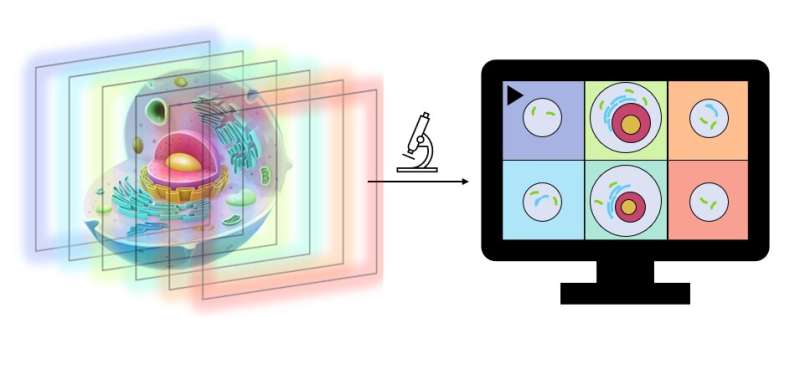
Although 3D microscopes already exist, their uses may be restricted by the slow pace at which the image is produced. These devices can typically only capture one to five photos per minute, which limits their usefulness for living and moving cells. In a typical 3D image, the depth of the forest can be seen, as well as which leaves and trees are closer together than others. The tiger hiding behind the bushes may be seen using the same technology as our new 3D microscope. One may view and examine many strata on your own, explained Florian.
Kenneth Bowitz Larsen, who oversees a sizable laboratory with cutting-edge microscopy equipment utilized by research teams at the Faculty of Health at UiT, tested the microscope. “The concept is brilliant; the microscope they have built does things that the commercial systems do not,” said Larsen. His laboratory mainly uses commercial microscopes from suppliers like Zeiss and Nikon. The laboratory also collaborates with research groups such as the one Florian represents. “They build microscopes and test optical concepts; they are in a way like the Formula 1 division of microscopy,” he added.
Why was this microscope created?
Although the commercial microscopes in Larsen’s laboratory must be able to do a range of jobs, the microscope created by Florian’s team is geared toward more focused activities. “It is very photosensitive and can depict the specimen in various focuses. It can work its way through the sample and you can view both high and low. And it happens so fast that it can practically be seen in real-time. It’s an extremely fast microscope,” said Larsen.

While the device created by Florian’s team is meant for more specific duties, the commercial microscopes at Larsen’s lab must be able to do a range of jobs. In addition to making it easier to see moving cells under the microscope, speed also helps to prevent photodamage. Cells are not fond of bright lights. Since this microscope operates so quickly, the cells are exposed to considerably shorter illumination, making it milder, according to Larsen.
The team applied for a patent and is looking for industrial partners to develop the microscope into a commercial product. To that end, the team is working to develop an upgraded version to make it easier to use for a broader audience.
The study was published in the journal Optica.
To ‘science-up’ your social media feed, follow us on Facebook, Twitter, or Instagram!
Follow us on Medium!