Home » The modern day Leeuwenhoeks

The modern day Leeuwenhoeks

by Editor CTS
English: Anton van Leeuwenhoek postage stamps ...
 Anton van Leeuwenhoek postage stamps Netherlands 1937
(Photo credit: Wikipedia)
When Anton van Leeuwenhoek invented the microscope, working out of his own shop in the Netherlands, little did he know the scale of the world, he was opening doors to. From being able to see little animalcules, as Anton called them in his letters to the Royal Society, we moved to a world, where we could isolate these microorganisms and study them. As our understanding improved, we started using these micro organisms for our own benefits like making cheese or beer and today, we know them so well, that we can edit their genetic properties to make vaccines and medicines for us. Yet, these tiny little beings continue their mischievous behaviour as they used to hundreds of years ago. In spite of all the antibiotics that we have, these micro-organisms can cause havoc in our bodies at the slightest opportunity available. The recent of case of Ebola in West Africa or the pandemic of AIDS are just examples of how we are still struggling to stay on top of this human-microbial see-saw. 
As the field of microbiology continues to grow, scientists discover new types of micro organisms, some useful while others harmful. Rather, adventurous scientists like Craig Venter have gone ahead and created synthetic life forms  on their own. While this might sound like a story of a sci-fi movie, with the crazy scientist trying to build a super bug, the larger scare is actually from the organisms that we know are harmful and are hiding amongst us, looking for that opportunity to strike. Tropical diseases have long been neglected by pharmaceutical companies, primarily, because there is little of hope of recovering research and developmental costs from sale of drugs in these countries. But now that we know that these micro organisms are capable of travelling across continents, governments are taking precautionary measures to avoid epidemics from occurring in first place. 
One effective way of preventing epidemics is containing the disease at the source. This is why international organisations such as WHO, FIND, MSF etc. have been working at grass root levels so that they can arrest the spread of such diseases before they turn into epidemics. Unfortunately, diagnostic methods in these remote areas are quite primitive. Just like pharma multinationals, companies dealing with new age diagnostic methods shy away from remote areas and prefer wealthy urban areas as their starting points for on-field implementation. There are some philanthropic organisations that try to avoid such neglect of the needy areas but such efforts are usually time consuming. The best way to address such issues is to to develop new techniques with easily available existing tools so that the costs of R&D remain low and even the remote areas can access them easily. One such effort is the CellScope, being developed by the Fletcher Lab at UC Berkley.  
The CellScope is a modern adaptation of the microscope that is aimed to be used by anybody and everybody. Breaking the tradition that microscopes belong to the lab and to the scientists therein, CellScope is a simple device that fits your pocket and can be carried everywhere. And why not? It is actually your smartphone, improved for scientific pursuit. There are many among us who boast about the capabilities of their new smartphones and probably, even the quality of ‘selfies’ that it can take. The Fletcher Lab is putting this ‘self-obsessiveness’ that smartphone companies use to sell us new models, to some good use by harnessing the power of their cameras for investigative purposes. 
Basic CellScope
Image credit: www.cellscope.berkeley.edu
Among the initial models built by the lab, is a platform consisting of comparatively higher optics, a source of illumination for the object to be studied and some custom software that can be applied to get standard (black and white) as well as fluorescent (coloured) images from a regular smartphone. 
Such a simple and small set up is easily portable and is also not demanding in terms of power or technical expertise. The team has successfully conducted some trials in countries such as Vietnam, Thailand, Cameroon, Ivory Coast etc. to name a few. 

This simple device can be used to detect cancerous cells in patient samples or simply identify the flora and fauna in water bodies. You could be in the middle of a sea, take a small sample of water and start recording your observations right on the deck with a CellScope. That is how quick this device is. But that is not what is really meant for!

What the inventors of CellScope want to do is make the device more smaller and portable and even simpler to use. This drive to make the device simple to use led to development of the OtoScope to help you keep a track on your child’s ear infections.

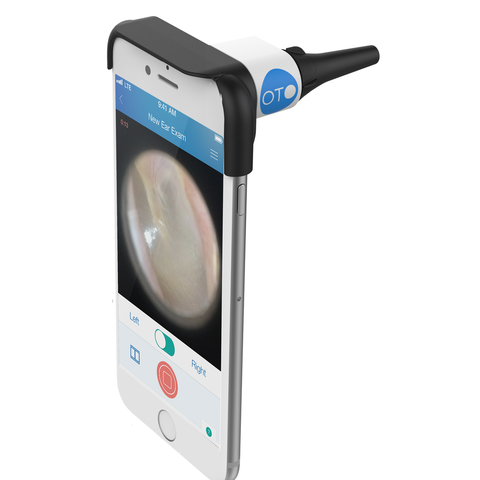
An Otoscope helps you take videos of the ear and send them to a doctor for his opinion. You get a diagnosis and even a prescription, if necessary, all within two hours, when you are still sitting in the comfort of your home.
Image credit: www.cellscope.com

A simple addition to your phone’s camera allows a parent to tackle ear infections at their first notice. Using the Otoscope, you can easily take a video of the inner ear and send it to a doctor, at any time of the day, without having to visit a hospital. The doctor’s team at Otoscope will review the video in less than 2 hours and send you their diagnosis and even a prescription where necessary. So, no more waiting at emergencies, just because you an innovative cellscope at your disposal.

But Cellscope is not just about making a device that saves you a hospital trip. Rather, recent developments on the CellScope have allowed researchers to come up with a life saving device. The work published in Science Translational Medicine, speaks about a pilot study carried out in Cameroon, where parasitic infections such as filariasis and onchocerciasis are major health problems.

Ivermectin, an anti-parasitic drug, is a simple treatment that can be offered to patients here, but this
drug administration has been compounded by another parasite called Loa Loa, whose presence in the blood, can lead to severe brain complications, if Ivermectin is administered to the patient. Identifying whether a person is infected with Loa Loa is a completely manual task and requires a highly trained technician and a conventional laboratory microscope. In such circumstances, the government’s efforts to carry out mass campaigns to inject Ivermectin and control filariasis and onchocerciasis, are severely affected, since arranging men and machine in remote locations for mass drives for the entire country is a logistical nightmare.

English: "Microfilaria of L. loa in a thi...
 “Microfilaria of L. loa in a thin blood smear, stained with Giemsa.” (Photo credit: Wikipedia)

An advanced adaption of the CellScope was used in the pilot project, where a single drop of blood was taken from the patient and loaded onto a capillary. The capillary is then inserted into an analytical base that is paired with a smartphone. All the user needs to do is inform a custom made app that the sample has been loaded after which, the smartphone communicates with the base, moves the sample near the phone’s camera and takes a video of the blood sample. The video is then analyzed by the smartphone for a wriggling motion that is peculiar to the Loa Loa parasite and then displays the number of worms spotted in the sample. All this analysis is completed within a matter of two minutes and requires no high end microscopy or even a well trained microbiologist to tell you where the nasty Loa Loa worm is present. Patients signing up for mass administration of Ivermectin can be quickly screened and protected against any adverse side effects. Called CellScope Loa, the device works something like this.

Aren’t these guys the modern day Leeuwenhoeks?

If you would like to know more about the Otoscope, CellScope or the CellScope Loa, you can find all the information at the Cellscope site at Berkeley University. For the readers interested in knowing more about the Loa Loa Cameroon Project, the link to the research paper is given below.

And if you would like to know more about such wonderful discoveries from the world of science, subscribe to our blog and we will send you an email every time we post something new and interesting. Alternatively, you can follow us on social media such as Facebook, Twitter or Google Plus!

Reference:

D’Ambrosio MV, Bakalar M, Bennuru S, Reber C, Skandarajah A, Nilsson L, Switz N, Kamgno J, Pion S, Boussinesq M, Nutman TB, & Fletcher DA (2015). Point-of-care quantification of blood-borne filarial parasites with a mobile phone microscope. Science translational medicine, 7 (286) PMID: 25947164

Related Articles

Leave a Comment